Anatomage Table Vet
The Anatomage Table Vet is a digital dissection platform that offers comprehensive anatomy resources for veterinary medicine, animal science, and zoology. With its digital real-tissue animal cadavers and extensive pathology library, Table Vet aims to improve learning outcomes while setting technological standards for veterinary institutes.
Discover the unique features

Accurate
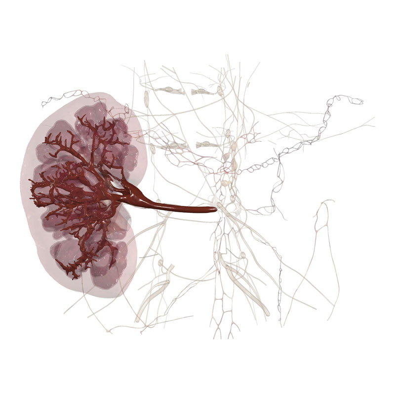
Rich content includes four real-tissue cadavers developed from actual animal bodies.

Ethical
Digital dissection eliminates the need for the use of living animals in veterinary education.

Extensive
1,185 segmented canine structures and 285 animal CT/MRI scans can be visualized in 3D.
World’s Most Canine Cadaver & Other Animal Tissues: Complement Anatomy Lectures with 3D Dissection
The Anatomage Table Vet creates an extraordinary learning experience for those who would like to study and interact with real animal cadavers in a chemical-free, ethically-inspired space. It offers digital versions of 4 real animal bodies, including a cat, mouse, frog, and the world’s most accurate and comprehensive digital map of a dog. With its highly-detailed animal cadavers, the Tablet Vet enables veterinary trainees to acquire real-life animal clinical experience right at schools.
Dissection
The Anatomage Table Vet allows for ethical dissection practices without the risk of harmful chemical exposure.
Veterinary Training
The tool facilitates hands-on learning for veterinary trainers through its 3D dissection capabilities, real-life animal cases, and detailed animal anatomy.
Animal Science & Pathology
CT/MRI scans, showcasing both normal and abnormal anatomy across various animal species on the Table Vet, provides invaluable insights into animal pathology.
Animal Anatomy Lecture Aids
Table Vet can connect to projectors, providing 3D visuals of animal anatomy and pathology, and fosters classroom collaboration by enabling content sharing via screenshots.